Google Janelia Maps Complete Male Fruit Fly Brain
Google Research and HHMI Janelia published a complete male fruit fly central nervous system connectome in Cell on 3 September 2026, mapping more than 166,000 neurons and roughly 125
PromptCrates Editorial
Staff Writer
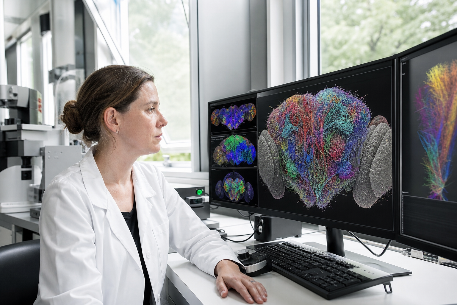
Google Research and HHMI Janelia published a complete male fruit fly central nervous system connectome in Cell on 3 September 2026, mapping more than 166,000 neurons and roughly 125 million synapses — the largest brain wiring diagram by neuron count to date. The atlas includes the ventral nerve cord and is viewable in Neuroglancer, giving labs a sex-comparable wiring resource for courtship, aggression, and sensory-motor circuits.
Why the male fly map matters now
Fruit flies already underpin Nobel-winning genetics work. Mapping their brains at synapse scale is the practical path toward understanding how nervous systems compute, because a human brain with about 86 billion neurons remains far beyond current reconstruction budgets. Google’s connectomics team and Janelia collaborators spent roughly a decade turning electron-microscope slices into verified three-dimensional reconstructions. The new male map sits beside earlier female brain and nerve-cord resources, so researchers can finally compare individuals and sexes in the same organism rather than guess from partial volumes.
The inclusion of the ventral nerve cord is especially consequential. That structure is roughly analogous to a spinal cord, so the dataset does not stop at perception inside the head. It begins to show how auditory, visual, and olfactory pathways drive motor outputs that control the body. For experimentalists, that means circuit hypotheses about walking, grooming, and courtship songs can be grounded in an end-to-end wiring diagram instead of pieced together from disconnected fragments.
Annotation and proofreading still required human experts at Janelia. Even with aggressive automation, error correction remains a multi-year bottleneck. The published resource is therefore not a raw AI dump; it is a curated atlas that labs can download, explore, and cite with confidence. Neuroglancer, Google’s open visualization stack for huge multidimensional volumes, is the primary portal for browsing the male connectome without shipping petabytes to every desktop.
AI flood filling and PATHFINDER progress
Connectomics pipelines start by slicing tissue into millions of thin sections, imaging each plane, and stitching those planes into a coherent volume. Google’s flood-filling networks grow from a seed pixel through convolutional predictions until they claim every voxel belonging to the same neuron. That approach powered earlier fully automated female reconstructions and the 2020 half-brain map with about 25,000 neurons and 21 million synapses. PATHFINDER, the team’s newer reconstruction system, improves speed and accuracy by mixing synthetic neurons into training data so the model sees harder edge cases before it meets real tissue.
Those engineering gains matter because verification, not imaging, dominates cost. Manual proofreading of neuron shapes can consume years of expert time. Techniques that cut that labor let the same labs attempt vertebrate brains that would otherwise stay out of reach. Google’s blog also notes companion methods for labeling and annotating neuron types, which reduces the chance that a beautiful mesh remains biologically opaque.
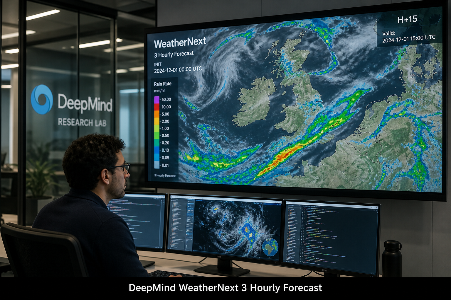
The male map already supports companion papers on visual systems, taste, and social behavior released alongside the Cell package. In other words, the resource is not waiting for a future user community; experimental groups are already running circuit analyses against it. That pattern mirrors how shared WeatherNext 3 forecasts moved quickly from research demo to operational testing once the underlying model weights and evaluation suites were public.
Fish brains and the road beyond insects
On the same week, a Columbia-led Nature paper used Google connectomics help to analyze a cerebellum-like circuit in the elephantnose fish hindbrain used for sensory prediction. The study is notable because it pairs a static connectome with plasticity and learning measurements, producing what the authors describe as one of the most complete mechanistic learning models yet available in a vertebrate. Larval zebrafish remain a parallel frontier: their brains are small enough for whole-brain mapping, and transparency lets experimenters record activity during behavior. Upcoming Harvard collaborations target whole-brain structure plus molecular type, while preliminary Fire&Wire-style datasets combine structure and electrical activity in the same specimen.
None of this claims a near-term human connectome. It does claim a ladder: flies today, fish next, selected mouse volumes after that. Each rung stress-tests flood filling, proofreading tooling, and visualization. Methods hardened on Drosophila then transfer to larger projects with fewer surprises. Healthcare ambitions remain long-range — better models of Alzheimer’s, depression, or schizophrenia circuitry — but the near-term payoff is experimental neuroscience that can ask causal questions with a shared wiring ground truth.
For AI practitioners outside biology, the story is also about evaluation discipline. Mapping brains rewards systems that admit uncertainty, request human review, and publish inspectable artifacts. That ethos sits close to debates about frontier lab disclosure after incidents like the German wiki agent swarm, where opaque internal tooling left outsiders reconstructing timelines after the fact. Open Neuroglancer volumes and Cell-level methods sections are the opposite pattern: publish the map, publish the pipeline, let others stress it.
What labs can do with both sexes mapped
Having male and female complete central nervous systems unlocks sexual dimorphism studies that partial maps could only hint at. Neurons that differ between sexes — for example projections present in male courtship pathways but absent in females — become measurable structures rather than cartoon sketches. In brain regions that look similar across sexes, two complete individuals finally let researchers quantify biological variability instead of treating one specimen as universal truth.
That dual-sex resource also clarifies motor planning. Courtship and aggression are classic Drosophila behaviors with strong genetic handles. Linking those behaviors to identified neurons across brain and nerve cord should accelerate optogenetic and silencing experiments that test causation, not just correlation. Pharmacy and medical imaging teams watching from the sidelines get a template for how AI-assisted reconstruction, human proofreading, and open viewers combine into a durable public good.
The Google–Janelia partnership frames the male connectome as infrastructure for a new experimental era. Success will be measured less by press headlines and more by whether hundreds of labs routinely cite the same Neuroglancer coordinates when they disagree about a circuit. If that happens, the 166,000-neuron milestone will look less like a trophy and more like the moment insect connectomics became routine scientific plumbing — the necessary precursor to vertebrate maps that actually resemble human-relevant computation.